


Thank you to Jimmy Sastra and Kitchener D. Wilson for discussions relating to this piece. All opinions are my own.
This essay is the first of three covering organoids. The full set is:
[Unreleased]
[Unreleased]
What are organoids anyway?
If someone placed a gun to the side of your temple and asked you to correctly define what exactly an “organoid” is, or else they’ll shoot, you may get a little nervous. The whole concept is something, you suddenly realize, you never quite understood. It’s more of a you-know-it-when-you-see-it thing, nobody in their right mind would ever ask for a clear definition, but you forgot to account for those who are not in their right mind. Desperately, you rack your brain for details. It’s an aggregate of cells, right? You stammer this out, tack on ‘it’s three-dimensional too!’, and stare at your antagonist expectantly. They consider your answer, and squeeze their hands.
If we were to interview your terrorizer later on, they would sheepishly admit that your answer is not the worst one they’ve heard. It really does encapsulate the many forms that organoids may take. Unfortunately, your given definition would also welcome blood clots, teratomas, and biofilms, all of which are indeed aggregates of cells arranged in three dimensions, but none of which would be considered an organoid by anybody on the planet.
You should not feel bad about this. In fact, you are in good company. The organoid field as a whole is not known to have particularly useful definitions.
One attempt at a definition comes from a 2014 Science review titled ‘Organogenesis in a dish: modeling development and disease using organoid technologies’, which defined an organoid as having the following traits: it is derived from either stem cells or organ progenitors, contains multiple organ-specific cell types, and recapitulates some type of organ-specific function. This feels more accurate, and swiftly removes blood clots, teratomas, and biofilms from the table.
Importantly, the organ-specific function bits of the definition help clarify something important: spheroids are not the same thing as an organoid. What is a spheroid? Here’s a useful infographic:
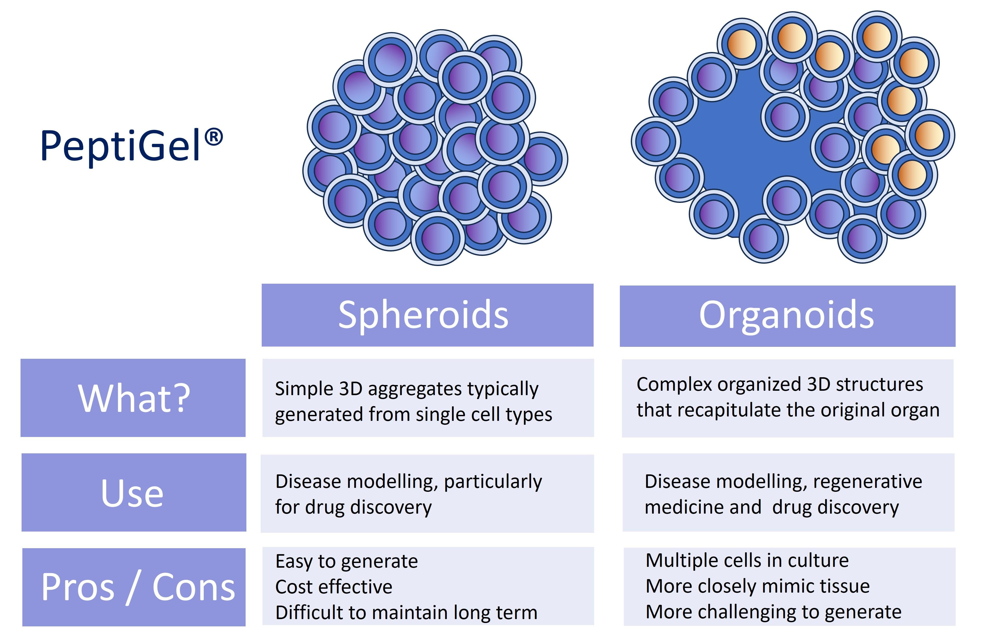
You, at the very start, described a spheroid.
Well, that’s that, right? Organoids are simply spatially complex, multicellular three-dimensional aggregates.
But we’re still missing some pieces. A 2023 Stem Cell Reports paper titled ‘Organoids are not organs: Sources of variation and misinformation in organoid biology’ has the following complaint about the particular definition stated above:
As a generic definition of an organoid, this fails to identify the distinctions between pluripotent stem cell [PSC] and tissue stem cell-derived [TSC] organoids.
The authors go on to say why this matters, but their explanation is dense and orthogonal enough to the point that we needn’t bother teasing out the details here, though we will later. What is important to understand is that PSC-derived organoids and TSC-derived organoids are doing fundamentally different things at the biological level, recapitulating different stages of tissue biology, and yet the word "organoid" is asked to contain both of them.
So: organoids are three-dimensional, multicellular, spatially organized, self-assembling structures derived from [cells] that recapitulate some aspect of [biology], though what cells and what biology will vary based on what the actual ‘organoid’ system is.
With regard to a definition, this is the best we can do. It is not very good, but it is good enough grounding to start discussing a very vital question.
Why haven’t organoids solved all of drug discovery?
There is a reasonable reaction one may have upon learning that organoids exist: why aren’t these being used all the time?
These are human, I repeat, human tissues that are lying in a dish, ones that are seemingly able to roughly organize themselves to semi-resemble complex, multicellular tissues. Why do we use mice, or any animal, at all? Hell, why do we even have clinical trials? Let’s just spin up a few hundred of these things and throw drugs at them. Get the FDA on the phone! It’s an emergency!
Yes, maybe the failure rate of this brave new endeavor will be high, but it’s a risk we should be willing to take. It’s not like we were doing so well before. After all, 99.6% of Alzheimer’s drugs fail, oncology programs entering Phase I succeed less than 5% of the time, and the average cost of bringing a single drug to market now regularly exceeds $2 billion, which is a number that has roughly doubled every nine years since 1950 and is so reliable it has its own law named after it. We’re terrible at this stuff. Why haven’t we switched to something that is clearly better?
It’s a good question! And there are a few very good answers.
The problems with organoids
The reproducibility of organoid work is (likely) abysmal
How do you make an organoid? The protocol varies by organ type, but the basic process is roughly the same. You start with cells, either stem cells derived from a patient biopsy (TSC approach) or pluripotent stem cells that you will coax toward a particular cell type (the PSC approach). You embed these cells in a three-dimensional matrix, usually in a gelatinous protein mixture called Matrigel, and then add in a cocktail of growth factors and small molecules that this particular cell type demands. Then you wait.
Over the course of days to weeks, the cells proliferate, migrate, sort themselves into different populations, and begin to self-organize into something that, under the microscope, looks like it might mirror the aforementioned micro-structures you are looking for. In some very real sense, this is a very accelerated and incomplete caricature of embryogenesis.
That’s the theory anyway. The problem with organoid creation starts with Matrigel—the stuff that most organoid globs are floating around in.
Matrigel is a gelatinous protein mixture secreted by Engelbreth-Holm-Swarm mouse sarcoma cells. Yes, cancer cells. And as you would expect would be the case with anything secreted by cancer cells, it is both extremely complicated and highly heterogeneous. Proteomic analysis has shown that the liquid contains 1,800 proteins, most of which we don’t understand, which can vary from batch to batch.
Surely the company behind Matrigel, Corning, would try to standardize things, no? Corning's solution to the batch variability problem has been a sort of radical transparency, in which they offer the aggregate mass of proteins in a particular Matrigel—ranging from 8 to 22 mg/mL—but no information as to whether specific proteins, say laminin, are more or less prevalent. I can’t find any work that studies how much this quantitatively hurts reproducibility—given that every lab is running a slightly different experiment depending on which Matrigel lot they received—but I can find a lot of papers complaining about it (here and here), so I’m going to assume it is a problem.
Why can’t we just stop using it? There are alternative options on the table that are in the works: decellularized extracellular matrix, synthetic hydrogels, and gel-forming recombinant proteins. Some of these work well, sometimes, for specific tissue types, but none of them are as versatile and easy-to-use as Matrigel. Another funny reason for lack of adoption is that validating these alternatives typically requires a reference point, and that reference point is almost always Matrigel-based.
The other layer of variation is upstream: the cells that are within the Matrigel. Here, there are so many axes of variation that it would actually be easier to just explain the empirical reality observed by one paper: ‘Cross-site reproducibility of human cortical (hCO) organoids reveals consistent cell type composition and architecture’.
Here is what they did:
To test hCO reproducibility, three IDDRC sites (Children’s Hospital of Philadelphia [CHOP], Children’s National Hospital [CN], and the University of North Carolina at Chapel Hill [UNC]) chose a widely cited guided protocol that demonstrated transcriptomic fidelity to primary fetal cortex and cortical wall-like organization.
…
The same iPSC line (PGP1), bioreactor parts, and Matrigel lot were used to standardize differentiations, allowing us to detect differences in hCO phenotypes due to handling differences across sites.
….
We assessed common phenotypes studied in hCOs, including cell type proportions, gene expression, and structure across time using several assays.
The good news is that some things stayed consistent: cell types and visual structural organization stayed roughly the same across the three sites.
The bad part is that the actual internals of the organoid varied dramatically between sites. To be specific: 786 unique differentially expressed genes (DEGs) across sites were detected at day 14 of the study, and by day 84 the total increased to 2,188 unique genes across cell types—most of which were cell stress or metabolic genes. Now, a DEG difference does not by itself establish functional difference, but it should make us a little wary.
What can we blame this on? We don’t know! Remember that the PSC line and Matrigel lot stayed constant, so those can’t be the culprit. The authors suggest a few ideas, but nothing is conclusively proved.
There is another interesting finding in the paper, though it doesn’t have to do with DEGs, but rather replicability amongst the organoids that the PSCs create. When one is designing an organoid experiment, it is good practice to ensure that the PSCs you’re working with are, in fact, both pluripotent and stem cells. This is typically done by assessing a small set of genetic markers. And one of the results from this cross-study paper is that the observed variability in cell-type proporation in the organoids is correlated with marker states that are not typically measured. Specifically, a set of markers that are associated with "primed" versus "naive" pluripotency; a spectrum that describes how far along the cells are toward committing to a particular developmental fate, even though both are still technically pluripotent. The problem isn’t that that primed-state variation exists! It is that primed-state variation exists amongst the starting substrate of an organoid, empirically leads to differences in organoid structure, and is not part of the typical quality control process.
But it’s not just primed-state variation! So many things can differ across organoids.
Consider the conference paper ‘Human iPSC derived organoid models to study tau pathology’, which found that different cell lines express the receptor TGFBR1/ALK5 at different levels. This is concerning, because SB431542, the inhibitor that at least a few cortical organoid protocols use to push cells toward a particular neural fate, acts on that receptor. If your cell line expresses more of it, the same dose of inhibitor does less. They fixed it in this paper by systematically measuring the receptor and adjusted the inhibitor concentration to match, which is fine work, but consider their explanations:
Well‐patterned organoids included 16 neural subtypes identified by scRNA‐seq, abundant rosettes, and robust BCL11B+/TBR1+ cortical neurons at 2 months.
In contrast, poorly patterned organoids contained mesendoderm‐related cells, identifiable by negative QC marker COL1A2 and/or few cortical neurons.
Do you see the problem here? The failure was visible because it crossed readouts they happened to measure, and they optimized against the measurements that exposed it! There is no reason to believe that there aren’t many, many equivalent deviations that were simply not surfaced by the readouts the authors were concerned with. I realize one can get infinitely paranoid about stuff like this. At some point in your cellular measurement journey, you’ll need to stop. But surely one protein marker and a few aberrant cell types can’t be enough.
So, we have two independent papers identifying two completely unrelated sources of PSC-level variation. To be fair, not all organoids are PSC-based, some are tissue-derived stem cells (TSC) that actually sidestep some of this. If you start with an organoid derived from a tissue biopsy, the cells are lineage-restricted, and so are less prone to the kinds of random walk through developmental space that affect PSC-derived organoids.
But now you’ve switched the cell-level variability concerns for patient-level variability! Yes, this may not be a problem for genuine, n=1 experimentation, but is a problem when you’re doing stuff at larger scales, requiring you to collect cells from many donors, each of which may have their own genetic quirks that can dramatically alter the outcomes of your experiment.
But we should be optimistic in cases like this. What if this variation doesn’t actually matter for translational purposes? Yes, perhaps perfection is impossible and anything short of direct human dosing will fall short, but maybe we get close enough that it’s fine.
In the ideal world, I could offer you a paper that directly assesses how much organoid technical variability affects translational utility. Unfortunately, while papers on each individual side exist, the connection between the two does not, and is likely at least several years away. We simply don’t know the impact of anything discussed here. After all, it’s only in the past few years that the organoid literature has even started to grapple with the variability question.
Speaking of translational utility, another natural inquiry with organoids one may have is: how much do they actually capture human responses to drugs?
Many therapeutic knobs are not exposed in organoids, and it is difficult to add them in
I wrote a few months back about how nearly all drugs work by taking advantage of some underlying knob in your biology, a receptor, an enzyme, something you can grab and turn. A useful question to ask about any preclinical system, then, is: how many human-like knobs does it expose?
It varies. Happily, it varies in a relatively clean way, dividing along three axes: the knobs that live at the cell level, the tissue level, and the body level. Organoids are excellent at the first, okay at the second, and essentially absent at the third. We’ll take them in order.
At the cell level, organoids are strongest. They have genes, they have proteins, and many of these genes and proteins are identical to the ones we adult humans have. As a decent rule of thumb, any disease where the entire causal chain of biological action is contained within a single cell, organoids are at least as good as any other in vitro system. In fact, they may even be better, since organoids are graded on their resemblance to real human cells.
At the tissue level, organoids continue to be quite good, but the jagged frontier begins to loom.
The good part first: organoids are, almost universally, plump. This is an immense technological leap over their cell-plate cousins, which are flat layers and thus completely unable to model the reality that drugs have to physically penetrate aggregates of cells in order to work. This is, of course, limited, because despite them being plump, they still are small. Most are a few hundred micrometers across, which is to say, the size of a grain of sand. The biggest ones get to a few millimeters, at which point they graduate to being visible to the naked eye, but remain specks. What does the speck look like? Depends! It might be a hollow sphere. It might be a branching structure with finger-like protrusions.
This is a good opportunity for us to agree on a good mental image for an organoid. If you are picturing a tiny liver, stop. If you are picturing a tiny anything, stop. You are picturing a tiny, incredibly small, dark dot, and it is important to not upgrade this dot too far beyond how you’d view a single cell.
But what of the ‘organ’ part of organoid? Yes, these dots do have structure. But the meaning of this structure is often vastly overinflated. When someone tells you their intestinal organoid has “crypt-like structures,” they mean something frighteningly banal: the surface of the sphere has bubbled outward into little bumps, and if you stain the bumps with the right chemicals, it vaguely resembles the arrangement you’d find in a real intestinal crypt. Similarly, a brain organoid with “cortical layers” is a blob where, within the blob, zones of different cell types settle at different densities, and the progression from one zone to the next is reminiscent of the layers of a developing human cortex.
Do the bumps or layers mean anything? Do they function the same way real intestinal crypts or cortical layers do? The authors of organoid papers sagely repeat these questions in their papers, discuss the importance of answering them, and proceed to—most of the time—not answer them.
To be fair, these simplifications may genuinely be fine. Organization in biology often implies function; cells arranged in a crypt-like pattern are presumably doing crypt-like things. Sometimes they are! And sometimes they very much are not. Which is the entire problem with tissue-level knobs in organoids! The knob might be there, in roughly the right place, maybe even turning. But whether turning it produces the downstream effect that turning the real human version would produce is a separate empirical question that has to be re-asked, drug by drug, phenotype by phenotype. There is, as far as I can tell, no universal agreement for when organoids can be safely relied upon in a given setting. Well, outside of a few cases, which will be discussed in a future essay.
At the body level, the knobs are simply not there.
A real organ in a real body has flowing blood, resident immune cells, and N years of age attached to it. None of these three things are in standard organoids, and these problems will be the subject of the rest of the section.
This blood bit, or perfusion, is probably the organoid’s biggest sin. No organoid system, left to its own devices, has yet generated a functional vascular network. Why is this such a big problem? Two reasons. One, without blood, you are dependent on the natural diffusion of oxygen to keep cells alive, and oxygen does not diffuse very far. The practical limit is somewhere around 200–300 micrometers from the nearest surface. Any cells further from that limit start to starve, leading to a ‘necrotic core’ in an organoid. This is an issue because it not only limits the absolute size of the organoid, but also messes up the cellular state of cells next to the necrotic core. And two, without vasculature, you cannot really model the PK properties of drugs. In a patient, a drug goes through a great deal of steps to reach its final location, interacting with many metabolic and physical processes in your body. In an organoid, the concentration of drug that an in-vivo cell sees is largely determined by how much you added in to start with.
Are there ways around this? Yes, but it does require you to cheat a bit. Either you co-culture with endothelial cells and coax the endothelial cells into forming vessel-like structures (which they do, poorly, and usually around the outside of the organoid rather than through the middle of it), or you transplant the organoid into a living animal and let the host vasculature invade.
You may notice I did not have a glib comment about this second option. This is because it actually works.
A 2013 Nature paper on transplanted PSC-derived liver buds is the first instantiation of the idea: human liver buds were implanted into immunodeficient mice and, over 48 hours, began to connect to its hosts vasculature. It worked well, so well that you are probably wondering, as I did, what has happened to this line of work. People have been actively trying to make something of it. A 2017 Cell Reports paper developed a platform to mass-produce liver buds at clinically relevant scales. A 2017 Scientific Reports paper worked on a newer transplantation method, since the original transplant sites in mice are nowhere you would put tissue in a human. A 2025 Molecular Therapy paper introduced PET-trackable buds so the fate of transplanted tissue can be monitored in vivo, since the field still does not adequately understand what happens to these things after they are transplanted.
This is cool research! Unfortunately, as you may have picked up from the subjects of the papers, it is not meant for drug screening, it is meant for transplantation into a human with a damaged liver. It may work for that, but it will not work for high-throughput drug screening, because our vascularized organoid is now stuck inside a low-throughput animal. Could you dig them out and use them as a screening platform? Maybe! But I haven’t found anyone who has tried.
Moving on to the next big organoid problem: the immune system. Curiously, few laymen think about vascularization being the biggest deal in organoids, but everyone is aware of the (immune x organoid) issue. It’s the first thing they bring up! And for good reason: there is basically nothing about drug response that the immune system doesn’t touch. Any drug with immune-mediated toxicity, any drug whose efficacy depends on immune engagement, and any disease whose pathophysiology is inflammatory are off the table.
Once again: is this fixable?
Well, if you’re working with some sort of patient-derived tumor organoid model, as in, grabbing tumor cells from a patient so they can replicate on your plate, you may get some autologous immune cells for free! Unfortunately, these don’t spontaneously reproduce, and grow smaller and smaller in count as you expand your organoid set to enable high-throughput screening. Well, can’t you just buy some more and throw them in? Sure, you can buy a vial of ‘Primary Peripheral Blood Mononuclear Cells’ (PBMCs) pretty easily for the low cost of $460.00, and it should contain a natural mixture of most immune-related stuff: monocytes, lymphocytes, and more, all isolated from healthy human donors.
Unfortunately, pouring these into the organoid culture also presents some challenges.
First off, the media that keeps the organoid happy and the media that keeps the immune cells happy are actively opposed to one another. One 2024 review termed it the ‘compatibility problem’, and did not offer a fix. Clearly there has to be one—given that there is an obvious proof point of the two coexisting within a human—but I struggle to find much progress. The practical consequence is that most organoid-immune co-cultures have to finish within 72 hours, which is a tight deadline for anything that isn’t related to direct cellular toxicity.
There’s also the HLA problem. The genes encoding these unique proteins, which serve as ways for our immune system to investigate cells, constitute the most polymorphic region of the human genome, which is to say, our vendor-purchased PBMCs and our organoid functionally came from two unrelated people whose immune systems would reject each other on sight. They do so here too, and they will often do so in a way that has nothing to do with the drug you add in. You can fix this by HLA-matching (expensive), by using autologous PBMCs from the organoid donor (limited by how much blood you can draw), or by deriving immune cells from the same PSC line as the organoid (finicky).
Okay. I’ve thrown a lot of accusations at organoids, but all this is theoretical, no? Unlike the prior section, there has been a lot of work on how well organoid drug-screening captures patient response. And it does mostly mirror the arguments here. Organoids are useful when the drug works the way the organoid works (cell-autonomous target, direct killing/functional rescue) and predict badly when the drug works the way the organoid cannot (immune, vascular, systemic). We’ll save a deeper look at that for later.
Well, wait a minute. We can’t end here. I mentioned three issues with organoids: lack of vascularization, lack of an immune system, and inability to age. The last one is different enough that it deserves its own section.
An organoid’s relationship to age is strange
One thing you’ll often see in organoid papers is a limitations section that says something akin to: caution, the cells that make up this organoid are fetal and have all the limitations that fetal cells have. But what does this mean?
When most people hear the word “fetal,” they think of a small version of an adult. And technically, yes, a fetus is a developing human that hasn’t finished developing. But what does it mean for a cell to be fetal? What properties define fetalness?
There are two: one of them is developmental stage and the other is chronological age.
Let’s start with developmental age. A developmentally fetal cell is a cell that expresses the developmental program appropriate to its gestational age; e.g. the first trimester according to transcriptomic comparisons. This leaks over to a lot of things! For example: metabolism. The dominant oxidative enzyme class in humans is CYP3A, and the dominant isoform changes across development. In fetal liver, the dominant form is CYP3A7. In adult liver, it is CYP3A4. So if you are testing a drug whose metabolism depends on adult liver enzymes, and your liver organoid remains stuck somewhere closer to a fetal developmental program, what good was the experiment? Now the answer may very well be ‘fine, since CYP3A7 and CYP3A4 aren’t terribly different from one another’, but these details still need to be thought through! For what it is worth though, they are quite different.
Well…who cares? Maybe the specifics don’t matter much. Maybe we just wait for the organoid to grow up?
Astonishingly, this works at least somewhat. A 2021 Nature Neuroscience paper cultured human cortical organoids for over 250 days and found that they cross a fetal-to-postnatal transition on a timeline that matches what happens in an actual human infant. A very reasonable thing to do here is to point a crooked finger at the authors and scream, ‘but how can you tell the cells actually stop being fetal?’. They do have a reasonable answer. In this case, the transcriptome and the methylation pattern matched those of postnatal cells via an epigenetic clock—Horvath was on the author list—and, more persuasively, the authors checked a small number of specific molecular switches that are known to flip postnatally in humans rather than gradually.
To be clear though: postnatal does not mean fully mature. Why? A lot of papers seem afraid to claim genuine, bonafide maturity. This makes sense to me; to some degree, it’s kind of an unprovable statement in an in vitro environment. We’ll discuss this in just a paragraph, but as a brief aside: isn’t it weird that waiting works at all?
It’s not too weird. You could imagine that this tempo is set by something close to bulk biochemistry—how fast proteins are made and degraded—and that it therefore was not something you could obviously turn up with a small molecule or a transcription factor. Phrased cutely: the organoid takes nine months to become a newborn because we take nine months to become a newborn. But we live in the future, and the future is much stranger. A 2023 Science paper found that modulating mitochondrial metabolism can alter this maturation; crank it up in human neurons and they mature faster, damp it down in mouse neurons and they mature slower. A 2024 Nature paper also identifies an epigenetic barrier that gates the timing, with its tuning leading to either arrested or accelerated developmental maturation. So at least a few knobs here have been identified, and perhaps the era of ‘mature organoids in a day’ is just around the corner.
Two caveats before we move on to the chronological age bit.
One, while the lines of evidence regarding maturity are nice to have, nothing about any of them establishes that the day-250 organoid neuron is doing what a genuine postnatal neuron in vivo ought to be doing. To connect this back: this feels like why people are afraid to claim maturity, because unfortunately for us and the neuron, there is no trustworthy way to check in full. Yes, there are partial methods to assess neuron function, but a real mature neuron is in a very neuron-friendly environment amongst its neuron-kin where it is doing neuron-y things—all of which are missing in our organoid. Recapitulating this in full is tough, because we’re still figuring out what all three of these things entail!
Two, the grander points here go beyond organoids. Comparisons of 2D and 3D culture have not found major differences in how fast the developmental clock advances, which suggests that mimicking tissue architecture—at least in this simplified manner—is not sufficient to move it.
Moving on: chronological age is roughly what it sounds like. The cell is young. And being youthful is a very deep property from a cellular standpoint: telomeres are long, somatic mutations have not yet accumulated, the protein degradation machinery of the cell is actually functioning at full bore, and on and on. This is a problem. If you believe that lots of diseases are downstream of some fundamental property of chronological age—and in fact, many are—an organoid cannot hope to model that disease in any meaningful capacity.
And unlike developmental stages, outrunning this one is a bit difficult. We certainly cannot wait eighty years for a neuron to grow a neurodegeneration phenotype! And even if we could, cells in a dish over time start gaining their own, stranger forms of aging that are disconnected from aging phenotypes that their in-vivo cousins have (a factoid that, interestingly, comes from a 1976 paper!).
However, being clever here has paid off. For instance, you can overexpress progerin, the mutant protein associated with Hutchinson–Gilford progeria syndrome, and get something that looks aged. Consider the following quote:
[Overexpressing progerin] induced nuclear morphology abnormalities, loss of LAP2α expression, formation of DNA double-strand breaks (γH2AX), loss of heterochromatin markers (H3K9me3 and HP1γ), and increased mtROS. These progerin-induced features were indistinguishable from those observed in primary fibroblasts from aged donors.
Of course, the same problem as everywhere else in this essay applies. Progerin gets you the readouts of age on the markers somebody thought to measure, and there are almost certainly plenty of facets of aging that we do not even know how to measure. Looking up ‘hallmarks of aging’ on Google Scholar will treat you to thousands of papers from scientists tripping over themselves to find yet another measure of aging. There are a lot of things going on in a cell!
To be clear however, these particular sins of fetalness—developmental and chronological—primarily apply to PSC-derived organoids. How about TSC-derived organoids? Nothing discussed in this section pertains to them! They’ve already accrued some facets of aging by virtue of being in a human, and have developmentally committed to a cell lineage. Kosher!
Unfortunately, TSC-derived organoids cannot be used to model any pathophysiology that is poking into territory where there are no stem cells, or at least no easily accessible ones. So this rules out cardiac conditions entirely, and largely rules out neurological conditions because humans are often unwilling to give up their precious brain stem cells.
So we’ll need to engage with PSC-derived organoids for at least some things. Are we doomed? Does their chronically fetal state make them useless? The honest answer is that we don’t know. A cell carries out a great many processes, the impact that fetalness has on them runs the gamut from partially documented to entirely unknown, and the impact that has on clinical translation is even less known.
Conclusion, and what are organoids good for then?
To close this out: I have been quite harsh on organoids so far, and, just like all forms of hate, doing so has been both fun and corrosive to the soul. Some kindness is in order. Just like all of us, organoids do possess some innate goodness. Yes, all the complaints from earlier are true, but there is some organoid work out there worth paying attention to, and it’d be an immense mistake to write them off entirely.
How can you tell the difference? You must go on vibe. A good organoid paper treats you kindly, softly. It knows it has hurt you in the past. It must regain your trust. It walks on eggshells around you, tiptoeing around as it explains itself, where it’s been, where it plans to go, cooing away your anxieties each time they arise. In the end, you and it embrace, the organoid promising to never wound you again, and you tearfully admitting that it will, that that is its nature, but you are happy it is trying to do better. If this does not occur, you should be wary.
But we must keep trying to forgive. What is the alternative?
Untrustworthy animal studies? Unrealistic binding assays? Extraordinarily expensive human trials? Any pessimist about organoids must admit that all of these have their own pathologies. The organoid field, much as some may believe otherwise, was not cooked up by some malevolent idiot who wanted the life sciences to spend several decades wasting their time. This stuff came about for a reason. This really is a genuine attempt at an optimal trade-off between ‘accurate’ and ‘cheap’. There has to be something worth exploring here with regards to translational medicine, because if there isn’t, we’re in a pretty dire situation.
And credit where credit is due: the hard workers over at the organoid factory have been listening to whiny essays like this for the past several decades, and are trying to fix things. There is a lot we did not discuss in this essay, such as microfluidics systems to incorporate some degree of ‘continuous flow’ over organoids, or, in popular parlance, ‘organ on a chip’, allowing you to—amongst other things—model some semblance of PK properties of drugs. Some of these innovations really do fix the major pathologies that organoids have! But they are still methods in progress.
However, there is so much worth discussing about this subject that I plan to release two more organoid articles in the coming weeks.
One of them will walk through some of the examples where organoids have been used for unarguably useful things. None of them have dramatically altered medicine, at least not yet, but still, progress! And reasons to believe that a better world is possible.
And the other will be a case study into a particular disease area where we really have no choice but to use organoids. And they have been used here, continuously, for over a decade. Has it been fruitful? Not really, but I plan to make the argument that it is probably good to dial in on it further.
Seems likely the field has been influenced by animal organoid systems that have been enormously productive: embryos that develop externally (no pregnancy, no Eutherians) have been used to make organoids for over a century. You can do cut-and-paste embryology, where you dissect away part of one embryo and stick it on another anatomical location in a different one (https://thenode.biologists.com/forgotten-classics-cut-paste-embryology/research/, the classic Spemann-Mangold organizer work). You can also isolate pieces that develop on their own in entirety (Holtfreter did this with amphibians in the 1930s, later folks adapted the trick to high-throughput measurements https://pubmed.ncbi.nlm.nih.gov/28103240/).
There's an obvious through-line here to modern organoid work - it would be really great if you could reliably do this with human tissues!